fbpx

The Joint Clinic

Wrist Surgery

Wrist Arthroscopy

Arthroscopy utilizes a small camera and microinstruments that enable your surgeon to see inside the joint without making large incisions into the muscle and tissue. Similar to arthroscopy of bigger joints like Knee and Shoulder, Wrist arthroscopy is used to diagnose and treat problems inside the wrist joint. The instruments inserted in the wrist are between 2 to 3 mm in diameter

Arthroscopic surgery can be used to treat a number of conditions of the wrist.

  • Wrist pain. Arthroscopy may be used to diagnose wrist pain when other tests do not provide a clear diagnosis. Often, there is inflammation or cartilage / ligament damage after a wrist injury which results in unresolved pain. After the diagnosis is made, the condition can usually be treated arthroscopically in the same sitting
  • Ligament/TFCC tears. The TFCC or Triangular Fibrocartilage Complex is a cushioning structure within the wrist. A fall on an outstretched hand can tear ligaments, TFCC or both. The result is pain with movement or a clicking sensation. During arthroscopic surgery, the surgeon can repair the tears
  • Wrist fractures. Small fragments of bone may stay within the joint after a bone breaks (fractures). Wrist arthroscopy can remove these fragments, align the broken pieces of bone, and stabilize them
Wrist Arthroscopy Setup
Dr Gaurav Gupta Performing a Wrist Arthroscopy
Previous
Next

Carpal Tunnel Release

This is done in cases of persistent pain, tingling, or weakness due to median nerve compression in the carpal tunnel at the wrist. The pressure in the carpal tunnel is released by cutting the ligament which stretches across the arch of the carpal bones (small bones of the wrist) and forms the roof of the carpal tunnel. This is a quick 10 to 15-minute procedure typically done under local anesthetic as a day surgery using a small cut of 2 to 3 cm on the palm. Most patients experience immediate relief from the severe pain, but it takes few weeks to regain full grip strength and lost nerve function

Diagram of carpal tunnel release

Dequervain's Release

This surgery is required for severe cases of De Quervain’s disease which don’t improve after conservative treatment. It is typically done under local anesthetic with the patient awake, as a day care procedure. The inflamed and thickened sheath around the affected tendons is cut to release the painful tendon entrapment. Although significant pain relief is felt immediately, it takes a few weeks for the surgical scar to become completely painless

Picture of scar after Dequervain’s Release

Trapeziectomy & FCR Interposition Arthroplasty (Suspensionplasty)

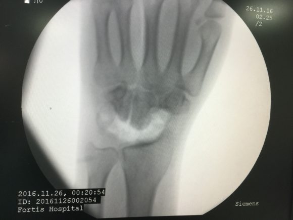
In persistent disabling pain due to arthritis at the base of the thumb, this surgical procedure is required. The Trapezium is a small bone in the wrist at the base of the thumb. This bone suffers wear and tear due to the arthritis, and is a source of pain. During surgery, this bone is removed to eliminate painful grinding at the thumb base. Since the loss of this bone can cause instability and weakness of thumb function in more active people, we sometimes fill up the gap left behind by using a rolled up ball of tendon harvested from the flexor carpi radialis (FCR) in the forearm

Proximal Row Carpectomy

Picture showing proximal carpal row removed
Scar-after-proximal-row-carpectomy-580x435
Scar after proximal row carpectomy

Proximal row carpectomy (PRC) is an effective procedure in the treatment of painful arthritis of the wrist. Many conditions— like scaphoid nonunions, scapholunate dissociation, kienbock’s disease can result in advanced collapse pattern of wrist arthritis. PRC converts the complex wrist link joint to a simple hinge joint. In this procedure, the surgeon removes three carpal bones in the row of bones that is closest to the forearm ie: removes the proximal carpal row. The distal carpal row (the second row of small bones) then comes down to fill the gap. The procedure is designed to reduce pain while maintaining wrist motion. Recently, more than 10 years follow up studies have emerged which have proved the safety and efficacy of this procedure

Wrist Joint Fusion (Arthrodesis)

Severe wrist arthritis causes disabling pain from the grinding of deformed wrist bones, apart from deformities and instability in the joint. A reliable treatment is to surgically ablate the wrist joint such that the forearm bones, wrist bones, and the hand bones form a continuous mass without any joint in between. The deformity is corrected at the time of the operation and the wrist is fixed in the functional position with plate and screws